

Copyrights: Tagwa Mohamed Ahmed Fadil, Mona Mohammed Hashim Ellaithi, Alkhair Abd Almahmoud Idris, 2024. License: This work is licensed under a Creative Commons Attribution 4.0 International License.
Abstract
Background: Breast cancer is recognized as a significant health issue in developed countries.
Methods: This study aimed to correlate the presence of Ag-NOR (argentophilic nucleolar organizer region) dots with immunohistochemical markers across various grades of breast ductal carcinoma. A total of 100 samples were divided into a test group and a control group, with the former comprising blocks of breast adenocarcinoma and the latter involving blocks of breast fibroadenoma. Each sample was sectioned twice; the first section was stained with Hematoxylin and Eosin (H&E), and the second utilized the Ag-NOR staining technique. The mean Ag-NOR (mAg-NOR) count and the proliferative Ag-NOR (pAg-NOR) count were calculated. Subsequent to microscopic examination of the sections, the laboratory findings and patient demographic data were analyzed.
Results: The majority of ductal carcinoma cases were classified as grade 2. The number of Ag-NOR dots was significantly greater in ductal carcinoma cases than in benign lesions. Furthermore, an increase in the number of Ag-NOR dots was observed with advancing tumor grade.
Conclusions: A strong correlation was identified between the count of Ag-NOR dots and the expression of immunohistochemical markers within different grades of breast ductal carcinoma.
Introduction
Breast cancer poses a significant health challenge in Sudan due to its high incidence rate1. The country's lack of comprehensive screening programs and diagnostic methodologies contributes to the ongoing prevalence of this disease among its population. Additionally, the high cost of diagnostic tests in Sudan places a financial strain on patients, making it difficult for many to afford the necessary care2. Consequently, there is a pressing need for the development of a simple, affordable, and accurate laboratory method for cancer diagnosis.
The protein Ag-NOR, associated with various biological traits of neoplastic cells—including their metabolic activity—is a potential marker for such diagnostic advancements. Ag-NOR proteins can often be identified using the silver nitrate staining method, a straightforward and rapid histochemical technique. This study aims to explore the utility of Ag-NOR staining in cancer diagnosis, particularly focusing on its effectiveness in determining cancer grades and thereby influencing treatment decisions and potentially enhancing survival rates through a cost-effective diagnostic approach.
Moreover, the study evaluates the long-term predictive value of the Ag-NOR staining technique in monitoring disease progression. There remains a gap in research concerning the integration of Ag-NOR staining into existing healthcare frameworks, specifically regarding personnel training, quality control maintenance, and handling the anticipated increase in diagnostic demand.
This research specifically targets Sudanese women and is conducted within a singular hospital environment, thus reflecting factors exclusive to the Sudanese healthcare system, patient demographics, and tumor characteristics inherent in this population. Given Sudan's unique healthcare infrastructure, economic landscape, and potential genetic variations among its population, the study addresses a critical void.
The primary goal of this research is to establish a correlation between Ag-NOR dots and immunohistochemical markers across different grades of breast ductal carcinoma, aiming to illuminate the prognostic and diagnostic potentials of Ag-NOR expression in a context-specific manner to Sudan.
Methods
This retrospective case-control study aimed to evaluate the efficacy of Ag-NOR staining in diagnosing breast adenocarcinoma among Sudanese women. The study utilized samples acquired from the histopathology laboratory of Omdurman Teaching Hospital. It included archived paraffin-embedded breast tissue from 100 women, divided equally into case and control groups. Cases comprised tissue blocks diagnosed with breast adenocarcinoma, whereas controls contained blocks of breast fibroadenoma. The inclusion criteria were strictly limited to female breast tissues diagnosed with either breast adenocarcinoma or fibroadenoma. Any other breast lesions were excluded.
A robust simple random sampling technique was employed to ensure the study's population was appropriately represented. This technique facilitated the division of samples into case and control groups for testing with Ag-NOR (silver nitrate) staining on paraffin-embedded, formalin-fixed tissues.
The staining protocol involved cutting sections to a thickness of 3 microns, which were then deparaffinized and rehydrated through a graded alcohol series before staining. Slides were stained in a dimmed light setting for 45 minutes and subsequently dehydrated, cleared, and mounted. Ag-NOR dots were quantified using an Olympus CHT microscope (Optical.Co.Ltd, Japan)3, 4.
Two experienced cytopathologists independently evaluated the proliferation index based on the stained slides5. The study spanned the entire period during which patients attended the histopathology laboratory at Omdurman Teaching Hospital.
Statistical analyses were conducted using SPSS (Version 20), focusing on frequencies, means, and chi-square test values to explore the association between Ag-NOR dots and immunohistochemical markers across different grades of breast ductal carcinoma. A p-value of <0.05 was considered statistically significant. Mean Ag-NOR (mAg-NOR) and (pAg-NOR) were calculated. After examination of the section under microscope, the results of laboratory investigation as well as the demographic data from the patients was analyzed.1, 2, 3, 4, 5, 6
Ethical approval was secured from the Al Neelain University Ethical Research Committee, in line with the Declaration of Helsinki Principles. Consent for sample and data collection was obtained from hospital administration, recognizing the challenges associated with retrospective consent for archived samples. Patient confidentiality was rigorously maintained, with patient information used solely for scientific purposes. Participants provided written informed consent, endorsed by the ethical committee of the Faculty of Medical Laboratory Research Board, Al Neelain University, Sudan. Ethical clearance code: AL-RES/03-022-06, dated 2/4/2022.
Results
The study involved one hundred participants, evenly divided into case (n = 50) and control (n = 50) groups. The mean age for the case group was 53.74 years, in contrast to the control group's mean age of 22.88 years, with members of the control group ranging from 10 to 40 years old.
Geographically, participants from both the case and control groups originated from various cities across Sudan. A detailed analysis of their provinces of residence indicated that, within the case group, 43.1% hailed from Western provinces, followed by 27.5% from Central provinces, 13.7% from Northern states, 7.8% from Southern provinces, and 5.9% from Eastern provinces (Table 1). For the control group, 41.2% came from Central Sudan, with subsequent distributions being 25.5% from Northern and Western Sudan each, and 5.9% from Eastern Sudan.
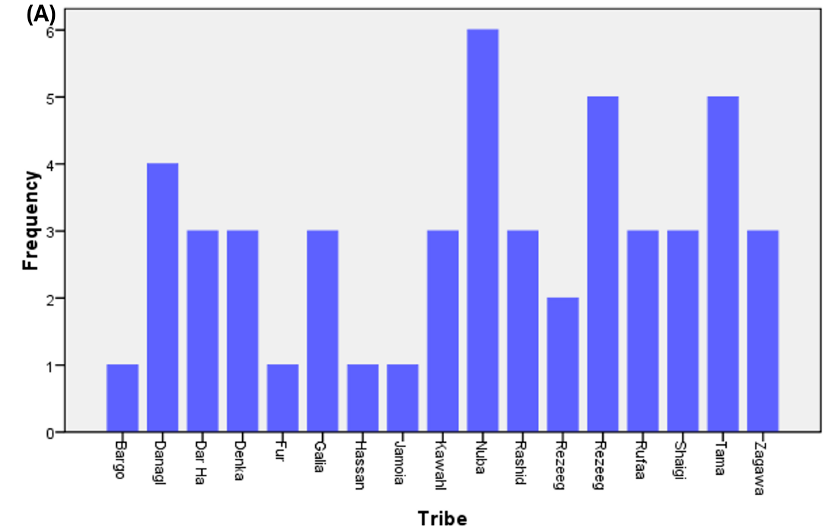
Additionally, the study categorized participants by tribal associations. The highest representation in the case group was observed from the Nuba tribe (11.8%), while the lowest (2%) included members from the Fur, Bargo, Hassnaia, and Jamoia tribes. Conversely, the control group displayed the highest representation from the Bargo tribe (23.7%) and the lowest from the Danagla tribe (2%). All participants were diagnosed with varying grades of adenocarcinoma of the breast, specifically, 16% with grade 1, 52% with grade 2, and 32% with grade 3 cancers.
The analysis extended to the activity of Ag-NORs on the slides of both the case and control groups, based on the count of dots per slide. In the case group, the counts ranged from 50–700 dots: 16% had 50–100 dots, 16% had 101–200 dots, 32% had 201–300 dots, 10% had 301–400 dots, 14% had 401–500 dots, 10% had 501–600 dots, and 2% had 601–700 dots. In the control group, dot counts ranged from 10–50, with distributions of 20 samples with 10–20 dots, 20 samples with 21–30 dots, 5 samples with 31–40 dots, and 5 samples with 41–50 dots.
Further analysis correlated the number of Ag-NOR dots with the grade of carcinoma. Among the grade 1 carcinomas, dot counts ranged between 50–100, whereas grade 2 carcinomas had 150–350 Ag-NOR dots, and grade 3 carcinomas had 400–600 dots.
Moreover, the study found significant differences in Ag-NORs counts in relation to other prognostic markers, such as nuclear loops, ER, and PR expression. Specifically, 50–100 Ag-NOR dots were associated with ER+/PR+ in 6 cases, 150–350 dots with ER-/PR- in 14 cases, and 400–600 dots with ER-/PR- in 8 cases (Table 2, Table 3).


| Location | Frequency | Percentage (%) |
|---|---|---|
| Centre | 14 | 27.5 |
| East | 3 | 5.9 |
| North | 7 | 13.7 |
| South | 4 | 7.8 |
| West | 22 | 43.1 |
| Total | 51 | 100.0 |
| Location | Frequency | Percentage (%) |
|---|---|---|
| Centre | 21 | 41.2 |
| East | 3 | 5.9 |
| North | 13 | 25.5 |
| West | 13 | 25.5 |
| Total | 51 | 100.0 |
| Grades | Frequency | Percentage (%) |
|---|---|---|
| Grade 1 | 8 | 16.0 |
| Grade 2 | 26 | 52.0 |
| Grade 3 | 16 | 32.0 |
| Total | 50 | 100.0 |
| Range | Number of cases | Percentage (%) |
|---|---|---|
| 50-100 | 8 | 16 |
| 101-200 | 8 | 16 |
| 2001-300 | 16 | 32 |
| 301-400 | 5 | 10 |
| 401-500 | 7 | 14 |
| 501-600 | 5 | 10 |
| 601-700 | 1 | 2 |
| Dots range | Number of cases |
|---|---|
| 10—20 | 20 |
| 21-30 | 20 |
| 31-40 | 5 |
| 41-50 | 5 |
| Grade | Ag-NOR number of dots | Cases number |
|---|---|---|
| Grade 1 | 50-100 | 8 |
| Grade 2 | 150-350 | 26 |
| Grade 3 | 400-600 | 16 |
| Number of dots Ag-NOR | ER + / PR + | ER + / PR - | ER - / PR + | ER - / PR - |
|---|---|---|---|---|
| 50-100 | 6 | 1 | 1 | 0 |
| 150-350 | 7 | 2 | 3 | 14 |
| 400-600 | 5 | 2 | 1 | 8 |
| Total | 21 | 5 | 5 | 22 |
| Grade/Marker | Grade 1 | Grade 2 | Grade 3 | Total |
|---|---|---|---|---|
| ER+/PR+ | 6 | 7 | 5 | 18 |
| ER+/PR- | 1 | 2 | 2 | 5 |
| ER-/PR+ | 1 | 3 | 1 | 5 |
| ER-/PR- | 0 | 14 | 8 | 22 |
| Cases number | 8 | 26 | 16 | 50 |

Discussion
The participant ages ranged from 30 to 80 years, with a mean of 53.74 years, aligning with findings reported by Adebamowo and Ajayi & Smith7, 8. A notable disparity was observed between the case group (mean age 53.74 years) and the control group (mean age 22.88 years), suggesting that age could significantly influence the study outcomes. Age-related variations in breast tissue characteristics may affect the expression of Ag-NOR dots and other immunohistochemical markers, potentially impacting result interpretation.
The study's inclusion of participants from diverse geographic and tribal backgrounds within Sudan could influence its findings. The representation from specific regions and tribes might introduce biases or limit the findings' generalizability to those areas only. Additionally, this diversity contributes to variations in genetic, lifestyle, and environmental factors that could affect the study outcomes. The case group primarily comprised individuals from the western states (43.1%), followed by the central (27.5%), northern (13.7%), southern (7.8%), and eastern states (5.9%). The control group showed the highest representation from the Bargo tribe (23.7%) and the lowest from the Danagla tribe, corroborating the findings of Khairy et al.9.
The distribution of cancer grades among participants (16% grade 1, 52% grade 2, and 32% grade 3) allows for a comprehensive analysis, with the predominance of grade 2 breast cancer reflecting specific diagnostic or disease patterns in the study population. Such a distribution might indicate an advanced disease presentation stage, common in areas with limited access to early detection and screening programs. This pattern aligns with various reports2, 10, 11, 12, 13, indicating a consistency with previous findings.
The variability in Ag-NOR dot counts between case (50-700 dots) and control (10-50 dots) groups highlights considerable variance in the biological behavior of breast adenocarcinoma and fibroadenoma, aiding in the differentiation between benign and malignant cases. The study emphasizes the cost-effectiveness of Ag-NOR staining in Sudan, proposing it as a viable diagnostic tool despite limitations such as staining variability and interpretation challenges that might affect reproducibility and reliability across settings.
An important observation is the association between Ag-NOR counts and immunohistochemical markers (ER/PR status), providing valuable insights into the hormonal receptor status of tumors. However, the study falls short of establishing causality and does not contemplate the integration of multiple diagnostic markers, which could enhance diagnostic and prognostic evaluations.
While the findings offer significant insights within the Sudanese context and suggest the potential of Ag-NOR staining as a cost-effective diagnostic method, the generalizability of these results to other settings necessitates further validation. The study points to areas requiring more research, particularly concerning the role of Ag-NOR dots in the progression of breast ductal carcinoma and the impact of cultural and social factors on adopting new diagnostic technologies in Sudan.
The study acknowledges limitations in exploring complex relationships due to its design and sample size, recommending these as focal points for future research. To validate the clinical utility of Ag-NOR staining more broadly, subsequent studies should aim for larger and more diverse populations, employ longitudinal designs, and integrate additional diagnostic markers.
Study Limitations
The generalizability of our findings may be constrained, primarily due to the study's focus on a specific population within Sudan, necessitating further validation to ensure applicability to other populations. The scope of this study does not encompass women diagnosed with breast cancer in preceding years, which may limit the comprehensiveness of our findings. The relatively small sample size, coupled with a lack of diversity in demographic and genetic backgrounds, suggests that our results may not be extendable to all populations affected by breast ductal carcinoma. Furthermore, the selection and range of immunohistochemical markers analyzed could potentially narrow the findings. Additionally, by excluding other breast lesions, the study's relevance to a broader array of differential diagnoses is diminished.
The sensitivity, specificity, and overall diagnostic accuracy of the Ag-NOR staining technique, along with its reproducibility compared to other methodologies, may impact the reliability of our findings. The variability in Ag-NOR dot counts observed across case and control groups could possibly reflect the method's sensitivity or specificity limitations.
Our study might not adequately reflect the dynamic nature of Ag-NOR dot numbers or the expression of immunohistochemical markers across different disease stages or in response to treatment modalities. Consequently, the ability of this study to provide broader insights into the prognostic significance of Ag-NOR counts and the correlation of immunohistochemical markers, such as ER/PR status, may be hindered by the limited sample size and study design.
Moreover, despite the involvement of two cytopathologists in interpreting Ag-NOR dots to reduce subjectivity, the reliance on human judgment cannot eliminate the potential for inter-observer variability, introducing another layer of limitation to our study.
Conclusions
The primary finding of this study indicates that Ag-NOR counts progressively increase from normal to benign, and subsequently to malignant lesions, highlighting the potential utility of this technique in distinguishing between these different tissue types. Ag-NOR scores are significant as they serve as an index to assess cell proliferation, which is pivotal in gauging tumor aggressiveness and predicting treatment outcomes. Furthermore, Ag-NOR scores enhance conventional histopathological analyses. They are utilized to discriminate between various grades of malignancy and can be integrated with routine histopathology as a diagnostic tool. The implementation of Ag-NOR staining has the potential to improve diagnostic accuracy or efficiency in histopathological laboratories, particularly in settings where resources are limited.
It was observed that the highest percentage of cases belonged to the Nuba tribe, with the majority of samples being diagnosed with grade 2 ductal carcinoma. However, the tribal or geographical prevalence presented in this study does not have clinical, social, or epidemiological implications. We advocate for additional research to investigate the utility of Ag-NOR staining in various types of cancer, its value as a prognostic marker, and its incorporation into standard diagnostic workflows. There is an evident need for future studies aimed at refining the application of Ag-NOR scores in clinical practice.
Abbreviations
Ag-NOR - Argentophilic Nucleolar Organizer Region, ER - Estrogen Receptor, H&E - Hematoxylin and Eosin, mAg-NOR - Mean Ag-NOR count, pAg-NOR - Proliferative Ag-NOR count, PR - Progesterone Receptor, SPSS - Statistical Package for the Social Sciences
Acknowledgments
Thanks for all participants involved in this research.
Author’s contributions
TMA and MMH conceived the design and carried out the experiments. AAI obtained, analyzed and interpreted the data. TMA and MMH wrote and revised the manuscript. AAI provides financial support for all experiments. All authors have critically reviewed and approved the final draft and are responsible for the content and similarity index of the manuscript. All authors read and approved the final manuscript.
Funding
None.
Availability of data and materials
Data and materials used and/or analyzed during the current study are available from the corresponding author on reasonable request.
Ethics approval and consent to participate
Ethical approval was obtained from AlNeelain University Ethical Research Committee in accordance with the Declaration of Helsinki Principles, and the agreement was taken from all hospital administration before sample and data collection. The patient’s information were highly secured and not used for other purposes than scientific inquiry. Each participant was asked to sign a written ethical consent form during the interview, before the specimen was taken. The informed ethical consent form was designed and approved by the ethical committee of the Faculty of Medical Laboratory Research Board, Al Neelain University-Sudan. Ethical clearance code number: AL-RES/03-022-06 Date: 2/4/2022.
Consent for publication
Not applicable.
Competing interests
The authors declare that they have no competing interests.
References
-
M.H. Bukhari,
S. Niazi,
S.A. Khan,
I. Hashmi,
S. Perveen,
S.S. Qureshi,
N.A. Chaudhry,
G.R. Qureshi,
M. Hasan,
Modified method of AgNOR staining for tissue and interpretation in histopathology. International journal of experimental pathology.
2007;
88
(1)
:
47-53
.
View Article Google Scholar -
Ahmed
H.G.,
Ali
A.S.,
Almobarak
A.O.,
Frequency of breast cancer among Sudanese patients with breast palpable lumps. Indian Journal of Cancer.
2010;
47
(1)
:
23-6
.
View Article PubMed Google Scholar -
Bancroft
J.D.,
Gamble
M.,
Theory and Practice of Histological TechniquesChurchill Livingstone Elsevier: Philadelphia; 2008.
Google Scholar -
Crocker
J.,
Boldy
D.A.,
Egan
M.J.,
How should we count AgNORS? Proposals for a standardized approach. The Journal of Pathology.
1989;
158
(3)
:
185-8
.
View Article PubMed Google Scholar -
Dasgupta
A.,
Ghosh
R.N.,
Sarkar
R.,
Laha
R.N.,
Ghosh
T.K.,
Mukherjee
C.,
Argyrophilic nucleolar organiser regions (AgNORs) in breast lesions. Journal of the Indian Medical Association.
1997;
95
(9)
:
492-4
.
PubMed Google Scholar -
Darkwaha
W.K.,
Gideon
A.,
Yanhui
A.,
Danquahc
K.O.,
Adjeic
E.,
Adankwahc
E.,
Assessment of proliferative index in different grades of breast cancers using AgNOR (Agyrophilic Nuclear Organizer Region) expression. Beni-Suef University Journal of Basic and Applied Sciences.
2018;
7
(4)
:
587-92
.
View Article Google Scholar -
Adebamowo
C.A.,
Ajayi
O.O.,
Breast cancer in Nigeria. West African Journal of Medicine.
2000;
19
(3)
:
179-91
.
PubMed Google Scholar -
Smith
R.A.,
Caleffi
M.,
Albert
U.S.,
Chen
T.H.,
Duffy
S.W.,
Franceschi
D.,
Global Summit Early Detection
Access to Care Panel
Breast cancer in limited-resource countries: early detection and access to care. The Breast Journal.
2006;
12
(s1)
:
16-26
.
View Article PubMed Google Scholar -
Khairy
G.A.,
Guraya
S.Y.,
Ahmed
M.E.,
Ahmed
M.A.,
Bilateral breast cancer. Incidence, diagnosis and histological patterns. Saudi Medical Journal.
2005;
26
(4)
:
612-5
.
PubMed Google Scholar -
Ploton
D.,
Menager
M.,
Jeannesson
P.,
Himber
G.,
Pigeon
F.,
Adnet
J.J.,
Improvement in the staining and in the visualization of the argyrophilic proteins of the nucleolar organizer region at the optical level. The Histochemical Journal.
1986;
18
(1)
:
5-14
.
View Article PubMed Google Scholar -
Eskelinen
M.J.,
Lipponen
P.K.,
Collan
Y.,
Syrjänen
K.J.,
The role of nucleolar organiser regions as prognostic factors in breast cancer. European Journal of Cancer (Oxford, England).
1991;
27
(8)
:
989-92
.
View Article PubMed Google Scholar -
Sujathan
K.,
Kannan
S.,
Pillai
K.R.,
Chandralekha
B.,
Amma
N.S.,
Nair
M.K.,
Significance of AgNOR count in differentiating malignant cells from reactive mesothelial cells in serous effusions. Acta Cytologica.
1996;
40
(4)
:
724-8
.
View Article PubMed Google Scholar -
Sofi
G.N.,
Sofi
J.N.,
Nadeem
R.,
Shiekh
R.Y.,
Khan
F.A.,
Sofi
A.A.,
Estrogen receptor and progesterone receptor status in breast cancer in relation to age, histological grade, size of lesion and lymph node involvement. Asian Pacific Journal of Cancer Prevention.
2012;
13
(10)
:
5047-52
.
View Article PubMed Google Scholar
Comments
Article Details
Volume & Issue : Vol 11 No 5 (2024)
Page No.: 6434-6441
Published on: 2024-05-31
Citations
Copyrights & License
This work is licensed under a Creative Commons Attribution 4.0 International License.
Search Panel
Pubmed
Google Scholar
Pubmed
Google Scholar
Pubmed
Search for this article in:
Google Scholar
Researchgate
- HTML viewed - 2805 times
- PDF downloaded - 883 times
- XML downloaded - 141 times
